Where Do We Go From Here: The Future of Hyped-up Dental Technologies
Considering how popular dental technologies may evolve can provide an exciting outlook on the future.
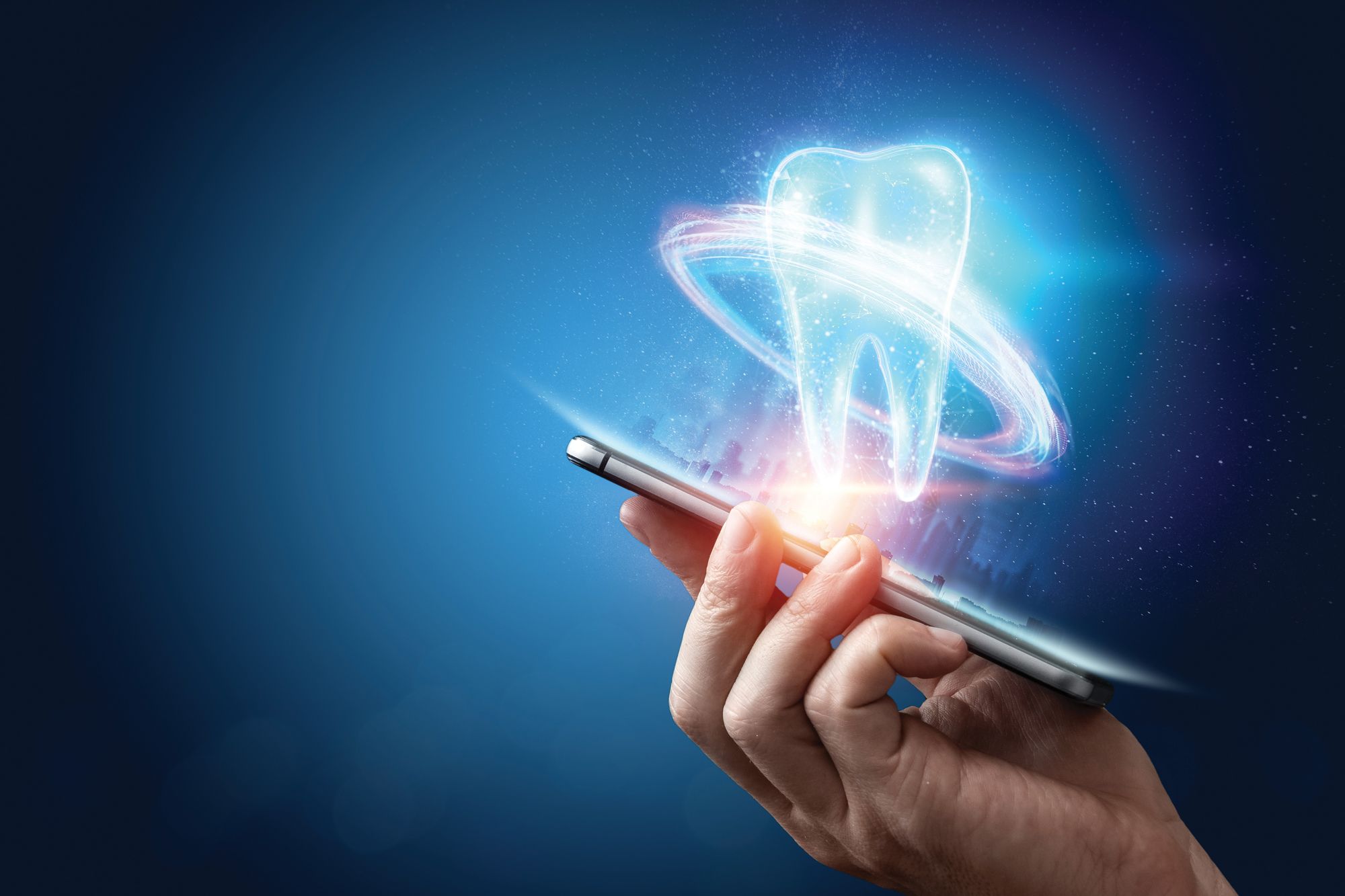
Where Do We Go From Here: The Future of Hyped-up Dental Technologies. Image courtesy of Aliaksandr Marko/stock.adobe.com.

When I was in dental school, we had procedural units we had to meet to graduate. One of those requirements was to perform 75 amalgam procedures. I find that kind of funny. We had students in a panic trying to finish up, needing to place 5 more amalgams so they could graduate. I look back now and realize I have not placed a single amalgam in the past 20 years. Crazy, isn’t it? Something I was required to learn isn’t even something I perform anymore. Honestly, I wish I had written down the last time I placed an alloy.
Don’t get me wrong; I am not against amalgams. If you are still placing the occasional amalgam, I understand why. Amalgam works. My patients, however, want esthetically pleasing restorations, and in my geographic area, composite is king. I still think it’s important that students be taught how to place amalgam. To know where you are going, you have to know where you came from.
One of the things I love most about our profession is how it continues to change and improve on a continuous basis. I’ve said it previously, but I’ll say it again: I rarely do anything the same way I was trained in school. I use many of the same principles (hence why I think learning to place amalgams is important), but those principles are applied in different ways with different devices and materials.
This month, we at Dental Products Report® decided to look at hype vs reality when it comes to dental technology. With that in mind, I’ve decided to retrieve my dusty crystal ball and discuss some topics for the future.
Visualizing 3 Dimensions
The idea of seeing things as they really are requires that we work in 3 dimensions. Dentistry has been heading in this direction for over 10 years, and that’s approximately how long it has been since cone beam computed tomography (CBCT) imaging began to go mainstream. Once computing power was hefty enough to process the data, CBCT use exploded in the profession. Many of us can’t imagine what it would be like to only work with 2D imaging now. Luxury becomes necessity.
This also applies to 3D intraoral scanning. This aspect of our industry is making incredible inroads. Many offices (mine included) now perform 3D scans for patients as part of a routine hygiene visit. The ability to see a patient’s mouth whenever you need to is a tremendous advantage to treatment planning. The number of impressions we take has dropped remarkably in the past few years, and that is mainly because cloud storage allows access to models at any time and any place we choose.
We are heading toward integrating all these data and merging them into a single, complete data set. Some of us are already able to merge intraoral scans into CBCT volumes. However, although it is possible in the current environment, it’s not common.
In the next few years, we are going to see what I call “the continually evolving 3D record.” Here’s how it will work: Every new patient will have a CBCT image taken and then have an image taken with an intraoral scanner. These procedures will be as much a part of the patient experience as completing a health history. The 2 files will then be merged so that the high-resolution intraoral scan is placed into the CBCT volume. Whenever new imaging is done, the new images will be integrated again.
The idea here is that anytime the patient has a new intraoral scan or a new CBCT scan, a corresponding integrated 3D record can be created. I envision the record-keeping process will not only track when something is changed by dentistry (such as a crown) but every time a patient has imaging performed. We all know that things change, shift, and wear in the mouth over time, and this will help us track that.
Many of today’s intraoral scanners have a time-lapse feature built in. This allows the patient to see a video of how their teeth have changed over time. I can see the same thing being done with merged 3D data to show patients how their oral health has improved or declined over time. The ability to communicate visually is a tremendous advantage of imaging, and better-informed patients make better health care decisions.
Artificial Intelligence
If you read my January 2023 article, you know our office just installed Pearl in January of this year. I am excited for what the future of this product holds. We are going to see incredible advancements using artificial intelligence (AI) in health care.
The study and discipline of AI has been getting better in fields outside of health care, where it didn’t need to help with critical and potentially life-changing decisions. Now that field of expertise has become robust enough to provide valuable information to help with diagnosis and potentially prognosis as well. Pearl currently has more US Food and Drug Administration clearances than any other dental AI product.
In the future, AI will not only provide data to help you make diagnostic decisions but may also suggest options for treatment. We’ve all had cases where you try to do what we affectionately refer to as “herodontics.” These are cases where a patient doesn’t want an extraction, root canal, or other treatment. So we try hard to meet those requests and expectations, only to be unsure of the long-term prognosis.
What if an objective third party could tell you something such as “there’s a 95% probability that this tooth will require endodontic therapy” or “80% of cases with this amount of coronal structure remaining report extraction and implant placement in less than 2 years”? Wouldn’t that be a great tool to have available to you? The opposite of this could happen as well, such as the system telling you that “you have treatment planned for an extraction, but 80% of cases with this amount of remaining coronal structure restored with a crown have a survival history of 5 years or more.”
I want to emphasize the point that the software will not make decisions. All it can do is provide more data for you to consider as you digest these data and make the appropriate treatment decisions for each individual case. You would consult the AI as you would a colleague who might sit down with you and offer suggestions on potential treatment plans.
Your Life in Pictures
Imaging isn’t just radiographs or intraoral scans. We can also help our patients make better decisions through extraoral photography. We are already seeing programs that can quickly and easily show patients their potential new smile. Take an extraoral photograph, and the new smile can be placed directly into the photograph. Although there are several programs on the market, one of the most impressive is exocad, which has Smile Creator. Smile Creator can show a patient’s new smile in a photograph and a 3D intraoral scan. This not only allows the patient to visualize the results but also helps the doctor design and create the needed restorations.
Let’s take this to a new level. In the not-too-distant future, you will be able to take a portrait photo with your digital single-lens reflex camera and then show your patients a complete makeover. Want to see how you would look with a new smile and green contact lenses instead of the blue eyes you were born with? Click! How about seeing how your appearance could be changed with Botox®? Click! Want to see how your lips would appear after dermal filler? Click! You get the idea. I could even see the potential for a photo booth in the reception area that allows patients to tinker with their appearance to see potential changes.
In-Office Milling
We are going to see incredible adaptation of milling soon, especially with the help of AI. Materials like zirconia and lithium disilicate are only going to improve, becoming stronger and more esthetic.
The design process will become remarkably faster and more accurate. Consider all the things I mentioned about AI in this column, and then imagine simply importing the STL file from a digital impression system and seeing the crown on the screen almost instantaneously.
If you desire, you will be able to change or improve the design so that it is exactly what you want. The system will assimilate those changes into its algorithm and learn your wants and needs. Over time, it will learn more and more about your individual preferences, suggesting restoration designs that are more in line with those preferences.
AI’s ability to design indirect restorations with ideal emergence angles, accurate margins, and spot-on occlusion is not science fiction. It is on the horizon and approaching rapidly.
Just for Fun
I won’t go into great detail on my ideas for this one, but since 2020 I’ve had a greater interest in infection control. We do our own fast, easy, and reliable sterilization monitoring in the office. So what about an autoclave that is preloaded with a sleeve of testing vials that run in each autoclave batch and is automatically transitioned to an incubator where it could be checked to ensure sterility is achieved every time the autoclave is run? I would love to see a system like that become available.
Wrapping Up
My thought process for this article was to look at technologies the industry is already familiar with and push them a little further. This article could be pages long. Things such as robotics, bioactive materials, and single-shade bulk-fill composites will continue to improve.
How lucky are we? This is the best profession on the planet. We are on an exciting and constantly evolving journey while getting to help patients and improve their quality of life. I am closer to the end of my career than I am to the beginning, but I am still enjoying the ride. Imagine what things will be like 5 years from now!

Product Bites – January 19, 2024
January 19th 2024Product Bites makes sure you don't miss the next innovation for your practice. This week's Product Bites podcast features new launches from Adravision, Formlabs, Owandy Radiology, Henry Schein Orthodontics, Dental Creations, and Dental Blue Box. [5 Minutes]
Product Bites – January 12, 2024
January 12th 2024The weekly new products podcast from Dental Products Report is back. With a quick look at all of the newest dental product launches, Product Bites makes sure you don't miss the next innovation for your practice. This week's Product Bites podcast features new launches from Videa Health and DentalXChange.