How to Revitalize Smiles With Advanced Zirconia Veneers
Navigating the challenges of modern veneer restoration leads to enhanced esthetic outcomes.
How to Revitalize Smiles With Advanced Zirconia Veneers. Image credit: © Linda Pearce, DDS

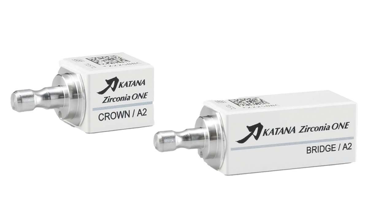
In the realm of modern cosmetic dentistry, the pursuit of optimal esthetics and durability often requires meticulous planning and the integration of innovative materials and techniques. This case study highlights the successful restoration of multiple zirconia veneers using the innovative KATANA™ Zirconia ONE layered zirconia (Kuraray Noritake). By leveraging digital technology, advanced materials, and evidence-based protocols, the clinician is able to address the patient’s concerns while delivering a functional and esthetically pleasing outcome that exceeds expectations.
Case Presentation
A woman with good overall oral health presented with concerns about the condition of her existing porcelain veneers on teeth #6 through #11. She reported a fracture on the lingual surfaces of veneers #8 and #9, as well as dissatisfaction with staining, black triangles, and lack of luster in the same area (Figure 1). The patient sought a comprehensive solution to restore the natural beauty and integrity of her smile.
Diagnosis and Treatment Planning
A thorough clinical assessment was conducted, including periodontal probing, occlusal evaluation, and diagnostic radiographs (Figure 2). Her periodontal health was found to be within normal limits, but the occlusion revealed a tight bite relationship with some misalignment of the opposing mandibular incisors, contributing to the breakage on the lingual surfaces of veneers #8 and #9. Radiographic examination revealed porous porcelain veneers #6 through #11 with microfractures throughout.
Based on the findings, 2 primary treatment options were considered:
- IPS e.max veneers (Ivoclar) made of lithium disilicate. While offering esthetic appeal, this option would require more extensive tooth reduction and provide less strength and longevity compared with the alternative.
- KATANA Zirconia ONE veneers made of layered zirconia. This innovative material, known for its beauty, strength, and longevity, emerged as the preferred choice due to its ability to address the patient’s concerns while minimizing tooth reduction.
Treatment Process
The treatment process began with a detailed digital impression using the CEREC Primescan (Dentsply Sirona) system’s BioCopy mode, which captured the existing porcelain veneers #6 through #11. This digital impression served as a blueprint for the new restorations, ensuring a seamless transition and maintaining the patient’s familiar smile esthetics.
Patient comfort and satisfaction were prioritized throughout treatment. Prior to the administration of local anesthesia, a cotton roll with lidocaine gel was applied for 5 minutes to enhance patient comfort. Buccal and interproximal infiltrations were performed using 2 carpules of lidocaine with 1:50,000 epinephrine. A putty impression was taken before the anesthetic administration to facilitate the fabrication of temporary veneers.
Next, the affected teeth were prepared for the new veneers. The existing porcelain veneers were carefully removed using a coarse diamond bur, and the teeth were prepared with a coarse chamfer bur, creating a wraparound “San Francisco–style” veneer preparation. A red fine diamond bur was used additionally to smooth the surfaces. This preparation design, with an upside-down smiley face on the lingual aspect, is preferred for its ability to conceal the margins in the anterior region, minimizing the risk of interproximal long-term staining. Opposing teeth #22 through #27 were also smoothed with a red fine diamond for optimal contours of teeth #6 through #11.
After the tooth preparations were completed, digital impressions were taken (Figure 3) and the margins for each veneer were outlined (Figure 4). A full-arch scan was performed using the CEREC Primescan, capturing both the upper and lower arches, along with an intraoral bite registration. This comprehensive digital scan provided the necessary data for the design phase. During the design phase, the CEREC Software’s proposal was meticulously adjusted to meet the case’s specific requirements. The BioCopy of the previous veneers was utilized as a starting point, with manual adjustments made to extend the incisal edges, narrow the interproximal black triangles using the shape tool, and set light contacts using the software’s form tool (Figure 5).
The material chosen for the restorations was the KATANA Zirconia ONE layered zirconia block in an A1 shade. Six blocks in size 14Z were used for teeth #6 through #11. This block size was selected for its ability to accommodate the dimensions of the anterior teeth effectively. The restorations were milled using the CEREC Primemill (Dentsply Sirona), with each block taking several minutes to mill at a fine speed setting. Subsequently, the milled restorations underwent sintering in the SpeedFire oven (Dentsply Sirona), following the recommended settings and firing cycles.
After sintering, the restorations were characterized and glazed. IPS e.max Glaze (Ivoclar) was applied using fine art brushes, with the inner portions of the veneers stuffed with a putty matrix and supported on a plastic stand. The glazing process was carried out in the CEREC SpeedFire oven, following the KATANA Zirconia ONE glazing mode parameters.
During the try-in phase, the fit and occlusion of the restorations were carefully assessed. The veneers were tried in pairs, and floss and articulation paper were used to ensure proper seating and occlusal harmony. For the final cementation, the intaglio surfaces of the veneers were sandblasted to enhance the adhesive bond, while the prepared tooth surfaces were etched and coated with CLEARFIL™ Universal Bond Quick (Kuraray) bonding agent. PANAVIA™ SA Cement Universal (Kuraray) in an A1 shade was applied to the veneers, which were then seated and spot-cured for initial stabilization. After removing excess cement and flossing, each veneer was cured for 40 seconds to ensure a secure bond. Occlusal adjustments were made as needed, ensuring a comfortable and balanced bite.
Challenges and Unique Aspects of the Case
One notable challenge during the treatment process was selecting the right shade for the restorations. With a considerably darker stump shade, a lighter block shade was chosen to offset the underlying darkness. Additionally, communicating with the dentist who performed the original veneer treatment proved beneficial in attaining the correct shade match. Achieving a seamless blend with adjacent porcelain restorations can be demanding, but the KATANA Zirconia ONE layered material excelled in this aspect, providing the best blend among available zirconia blocks for anterior restorations.
Treatment Outcome and Long-Term Follow-Up
Since completing the treatment, the patient’s oral health has remained excellent. A follow-up appointment was scheduled 1 week after the delivery appointment to assess occlusion and make any necessary adjustments or refinements to the incisal edges. The fabrication of a soft night guard may also be recommended at this appointment to protect the veneers from occlusal forces. Regular recall appointments with prophylaxis are recommended to maintain optimal oral health and prevent future issues. KATANA Zirconia ONE veneers provided a fresh, improved, and naturally beautiful appearance, restoring the patient’s confidence in her smile (Figure 6).
Key Learnings
This case highlights several key takeaways and recommendations for dental professionals. The layered zirconia material, combined with a digital workflow, offers a streamlined and efficient solution for delivering high-quality anterior restorations in a single clinical setting.
Careful shade selection, communication with previous treating dentists, and the use of customized shade guides can facilitate achieving a seamless blend with adjacent restorations.
The San Francisco–style veneer preparation design, with an upside-down smiley face on the lingual aspect, effectively concealed the majority of the margins in the anterior region, minimizing the risk of long-term staining.
Implementing advanced materials and techniques can enhance patient satisfaction, treatment outcomes, and the long-term viability of dental practices. Continuous education and a willingness to embrace modern technologies and materials are essential for dental professionals to stay at the forefront of their field and provide their patients with the best possible care.
Summary
This clinical case study exemplifies the remarkable outcomes that can be achieved through the synergistic integration of digital technology, innovative materials, and evidence-based protocols in cosmetic dentistry. By leveraging the exceptional properties of layered zirconia material and following a meticulous treatment approach, the clinician successfully restored the patient’s porcelain veneers, achieving a beautiful, functional, and long-lasting solution that exceeded expectations.
