The Maximum Advantages of Being Minimally Invasive
The way we practice minimally invasive dentistry and how we define it have changed greatly over dentistry’s history.
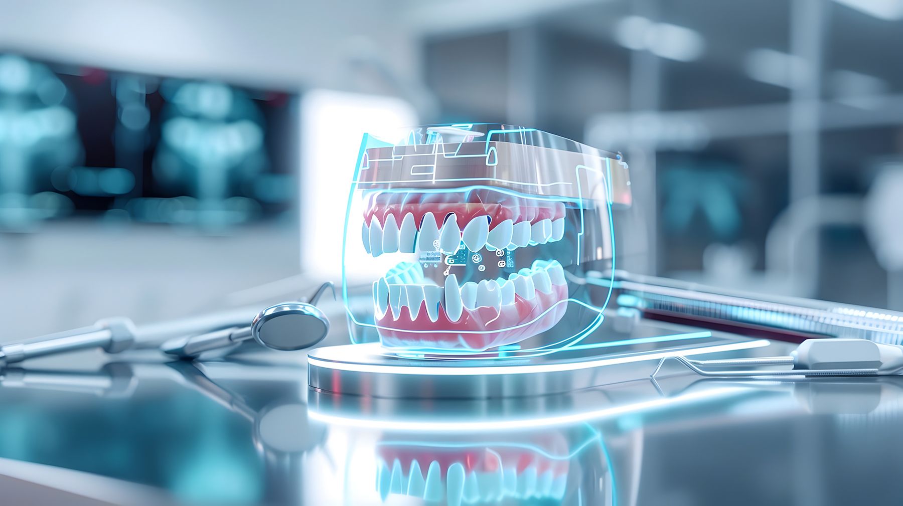
The Maximum Advantages of Being Minimally Invasive | Image Credit: © Phatsa - stock.adobe.com/Generated by AI

I’m back and ready to wrap up 2024 with this year-end issue of Dental Products Report. We focus this month on minimally invasive dentistry (MID), a topic the profession has been interested in for some time. We have probably been focused on being minimally invasive for as long as modern dentistry has been around—and by that, I mean for the last 150 years or so.
Some incredible advances in this concept have been made over the course of my career. During my days in dental school, the focus was on really well-done amalgams. Of course, amalgams were anything but minimally invasive, unless we are talking about a buccal pit. Amalgams needed a minimal width and a minimal depth to resist fracture. That meant we often sacrificed unaffected tooth structure just to get the prep to an adequate depth and width, frequently a very conservative 2-surface restoration fractured at the isthmus because the operator was trying to conserve tooth structure and there was insufficient bulk to resist the occlusal forces.
Amalgam doesn’t bond to dentin or enamel, which means it also requires mechanical retention. Doctors were forced to create undercuts on the occlusal and interproximal surfaces to create a retention form that essentially locked
the restoration.
I was also trained in the philosophy of “extension for prevention.” The basic idea was that if you were going to put bur to tooth, you might as well go into all the pits and fissures to include them in the prep. The thought was that as amalgam did not decay and teeth did, you might as well include areas of potential breakdown to the restoration to help prevent breakdown in the future.
The Early Days
Back when restorative dentistry began, MID had a different definition, probably because anesthesia did not exist yet. When you were excavating decay, you wanted to stay as far away from the pulp as possible simply because it hurt. A revolution occurred in the 1870s when actual dental-
specific handpieces were invented. However, there was still no local anesthesia. I feel this was when the extension-for-prevention idea became a real thing. If you had a chance to prevent further decay by being able to run out grooves, why not? In those days, people only sought treatment when in pain. If a patient was having a restoration done anyway, why not decrease the chance of future problems?
In 1884, the brilliant surgeon Dr William Stewart Halsted (chief of surgeons at Johns Hopkins University School of Medicine) read about the topical anesthetic properties of cocaine. This led him to conduct some experiments where he prepared a solution of cocaine powder and water that was placed in a syringe and used in various areas of the arms and legs to cause anesthesia. He then delivered it to a medical student volunteer. The result was the first successful anesthesia of the inferior alveolar nerve. This changed everything.
However, the profession was still far from having safe and predictable local anesthetics. Injecting cocaine into patients had many drawbacks. Of course, when it was first discovered, it was considered a wonder drug. The year 1904 saw the discovery of procaine (Novocaine), which worked without the problems of cocaine but was quickly metabolized, which meant a short duration of action. The real revolution in dental local anesthesia came in 1946 with the discovery of lidocaine. At long last, patients could be reliably anesthetized for an entire procedure.
Enter Composite
Then along came composite, and the world was rocked again. At long last, dentistry had a material that did not require mechanical retention. It flexed similarly to tooth structure, so bulk was not always needed for strength. Plus, it was beautiful.
Composite allowed doctors to be much more conservative than ever before. Dentistry went from extension for prevention to constriction with conviction, where the focus became on removing only diseased or weakened tooth structure. Gone were the days of 2-surface restorations dovetailing into the occlusal surface, replaced by the current philosophy of “slot preps,” where only the marginal ridge and interproximal caries are removed and replaced.
Enter Digital Diagnosis
Our decision-making process has also undergone major shifts. Bringing digital tools into the treatment sphere has allowed us to see things better than ever before. Almost everyone has readily available intraoral cameras, but other tools exist. Transillumination and fluorescence are also great ways to find caries at the earliest stages.
The Microlux DWfrom AdDent is a great, affordable device for transillumination and fluorescence detection. The device is a small handheld unit with a small tip (3-mm diameter) that can be easily used in both the interproximal and occlusal areas. It has 2 settings: 1 for white visible light for transillumination and 1 for purple (405 nm) that can be used to look for caries fluorescence.
KaVo also has a recently released device called the DIAGNOcam Vision Full HD—a sleek device that looks like an intraoral camera. However, it does much more. The device takes high-definition intraoral photos, transillumination (using infrared) photos, and fluorescence caries detection photos. The best part? You can take all
3 image types with the push of 1 button.
Advanced devices such as these can help us find problems when they are tiny and allow us to perform equally tiny restorations.
Enter Bioactive Materials
Now dentistry has moved into the realm of truly bioactive materials that can help the tooth rebuild itself. We all know that the area most susceptible to recurrent caries is at the restorative margin. I like to say that the margin is the trench warfare of dentistry. Because of that, I am excited to see manufacturers creating restorative materials that not only seal the margin but contain components that can fight recurrent decay and even remineralize weakened structure. Another great thing about these products is that although some are fairly recent additions, they contain components with a long track record.
As an example, I will use the RE-GEN™ product line from Vista Apex. This product line contains Bioglass, which was discovered in the late 1960s and has been used in orthopedic surgery for decades. Without going into excessive detail, Bioglass helps create hydroxyapatite where it contacts the tooth. That means it helps seal margins and helps the tooth repair itself under restorations. It also creates an alkaline environment along the margins, which has bactericidal effects.
Bioglass is currently available in the RE-GEN product line, which includes a bonding agent, a pit and fissure sealant, a flowable composite, a bulk fill composite, and an endodontic sealer.
Then there is GIOMER technology from Shofu Dental. GIOMER has a history dating back about 20 years and is present in many Shofu products. It is a bioactive surface prereacted glass that releases fluoride and 5 other beneficial ions, and one nice feature is that it also recharges them. That means that there is an active effect. GIOMER is a part of many of Shofu’s products, including universal composites (Beautifil II and Beautifil II LS), specialty composites (Beautifil II Gingiva and Beautifil II Enamel), flowable composite (Beautifil Flow), and more.
Another product line with bioactivity is ACTIVA™ BioACTIVE from Pulpdent. These products contain a product called Embrace Resin Cement. The unique aspect is that Embrace resin contains a small amount of water as well as phosphate acid groups. In the oral environment, this material loses hydrogen ions, which are replaced by calcium ions from the tooth; this forms a resin-hydroxyapatite complex and helps seal the restoration to prevent microleakage. It also absorbs ions from the saliva to help recharge its bioactive properties.
Going Deeper
Bioactivity also can promote healing and aid reparative dentin in areas where caries approach the pulp. Usually we think of minimally invasive as being tiny preparations with tiny restorations, but we can apply bioactive materials in areas of near pulpal exposures for large restorations, even full coverage.
When deep decay approaches the pulp, reparative dentin formation can help prevent sensitivity and help the tooth recover from the mechanical insult of caries removal. In these situations, the go-to has always been calcium hydroxide. However, new materials have put a new spin on this.
BISCO’s TheraCal® LC is a resin-modified calcium silicate material. It is designed to be used in direct and indirect pulp capping procedures and as a general protective liner. Additionally, it releases calcium and helps create an alkaline environment to promote healing. The material assists with hydroxyapatite crystal formation. Plus it is light cured, which ensures the set and reduces the amount of time required for the procedure.
There is also Ultra-Blend™ plus from Ultradent. This material contains calcium hydroxide in a urethane dimethacrylate base. It is light activated, and the resin is highly filled, which means it experiences minimal shrinkage as it polymerizes.
Wrapping Up
The profession has seen some amazing advances in materials in the past decade. During the early explosion of bonded restorations, doctors’ greatest concerns were polymerization shrinkage and occlusal wear. The material scientists who make our jobs easier have more or less solved those problems. We are now moving into materials that continue to work long after the patient leaves our office.
Study data show that many composites fail within 5 years, and that failure has been mainly due to the problem of recurrent decay at the margin. However, we are now seeing materials that can better seal the margins, help regenerate the needed mineral matrix, and even kill bacteria at the tooth or restoration interface. Beauty is a terrific quality, but beauty anddurability in the challenging world of the oral environment are what we all strive for daily. We are now seeing materials that are helping make that goal a reality. What a great time to be in dentistry.
