Digital Radiography Troubleshooting
Various digital radiography challenges due to factors like x-ray heads, sensors, and exposure times can affect practice performance. By applying a differential diagnosis approach, practices can find the solution and improve their image quality without breaking the bank.


Digital Radiography Troubleshooting. Image credit: © Ivan – stock.adobe.com
Although many of my articles focus on cybersecurity and Health Insurance Portability and Accountability Act compliance, I am asked to assist offices with many other things. Having spent 10 years as a periodontist, I am uniquely positioned to work with practices in areas in which other information technology companies have little experience. I recently visited an office in Pennsylvania to help diagnose less-than-satisfactory digital x-rays, and I thought how I handled the troubleshooting would make a good article.
One of the main challenges in digital radiography is that many variables exist between the taking of the image and what appears on the screen. In no specific order, these include the x-ray head, the sensor, the patient, the USB cables, the monitor, the video card…the list goes on and on. When the practice first contacted me, the initial thought was that the images were diagnostic but that there were just some people in the practice who could not differentiate between a good film image and a good digital image (digital x-rays tend to be a bit softer or fuzzier).
When I arrived onsite, I realized a few things:
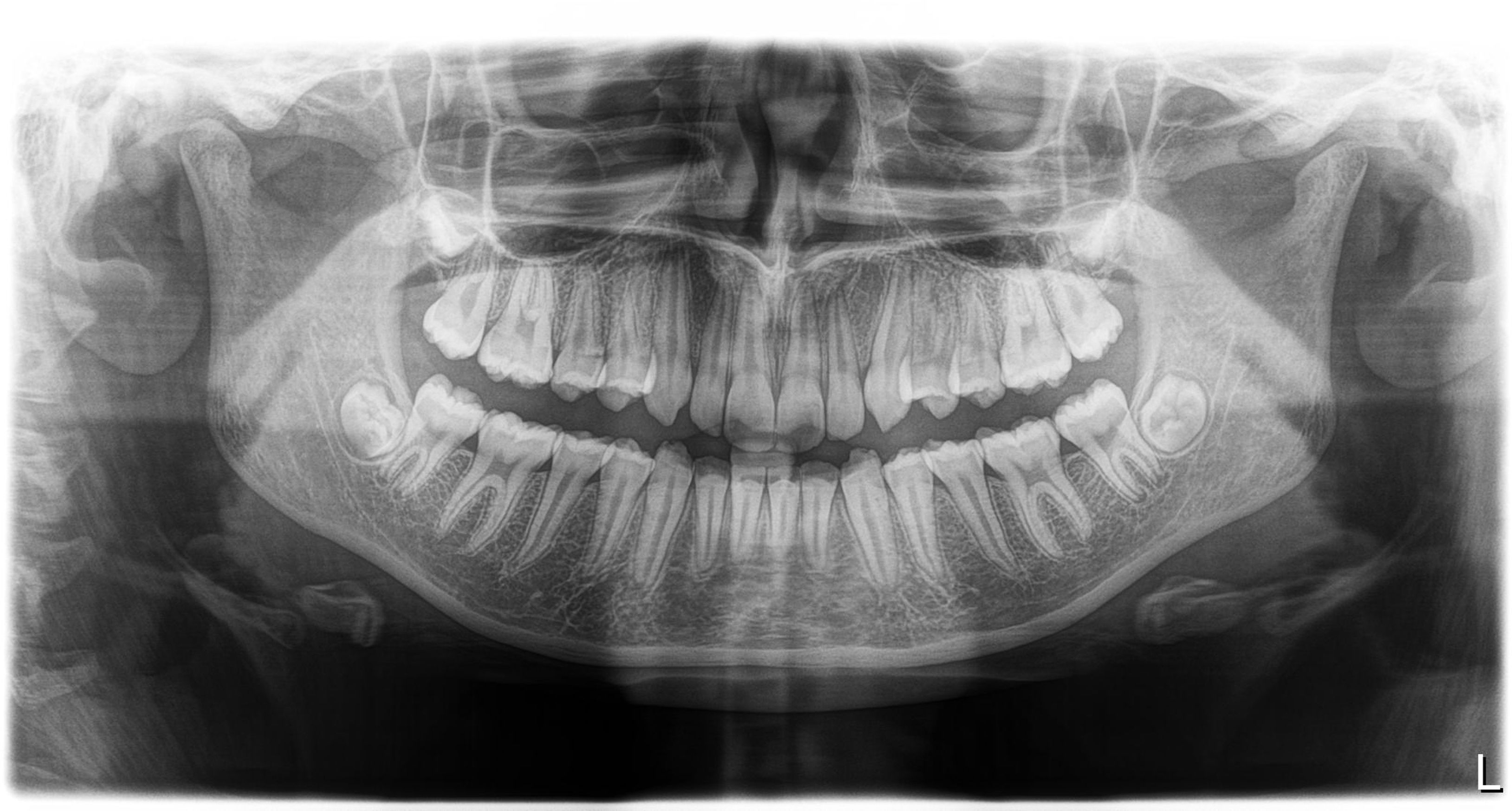
The images were indeed not diagnostic. There was considerable graininess in the images, which is almost always due to underexposure.
As I evaluated the setup in more detail, I saw that every image had a sharpening filter applied, which tends to accentuate the graininess. When I removed the filter, the raw image was completely undiagnostic; it was not capturing enough information.
When I visited a second location for this group practice that used the same software and settings but different sensors and x-ray heads, the images were far more diagnostic.
So, what conclusions could I draw, and how would I test them?
The primary way to troubleshoot is to do a differential diagnosis. I had to determine the most likely diagnosis and work from there.
The most common culprits are the x-ray heads and sensors.
Software and computer infrastructure can also cause similar issues, but because both locations had the same computers and monitors, I ruled those out as the leading causes.
One thing I suggested, and we tested, was bumping up the exposure times. Back in the day, digital x-rays required less radiation than film, so many people still have this too low. The practice used 0.16 seconds, but when I tested at
0.20 and 0.25 seconds, there was no evidence of overexposure. I suggested they keep going higher until it was clear that the exposure was too high, then step back 1 setting from there.
I suggested that they take the x-ray head from the office with good x-rays and test it at the office with bad images; it is easy to do because they have a portable NOMAD handheld system (DEXIS).
I also suggested they take a sensor from the good office and test it at the office that needed to be getting better images.
Anytime you deal with systems with multiple steps in their processes, it can be challenging to isolate the one variable causing the issues. Approaching it just like we approach patients with nonspecific pain is often the best course of action to determine the problem and how to fix it.