The art of smile design
Earlier this year, I competed in the first North American Candulor KunstZahnWerk® “Art of Denture” competition featuring Candulor’s “Swiss art of Prosthetics” and Camlog’s “perfect fit” implants.
Earlier this year, I competed in the first North American Candulor KunstZahnWerk® “Art of Denture” competition featuring Candulor’s “Swiss art of Prosthetics” and Camlog’s “perfect fit” implants.
The primary assignment was to complete a maxillary full denture and a removable mandibular bar retained full denture using the Gerber lingualized set-up method. The second part of the assignment was to document the procedures used to produce the submitted design for the competition.
I received third place overall and second for digital presentation.
Every participant received the same case and duplicated models to complete the prosthetics. The patient was a 72-year-old female who had worn dentures for 28 years. She suffered from poor fitting dentures and complained about problems with her speech, chewing, sore spots and the unnatural look of her anterior teeth. Upon radiographic examination, four Camlog implants were placed in the anterior mandibular bone.
The case was articulated to an average value with the right condylar inclination set to 32° and the left condylar inclination set to 36°. The anterior set-up was set in a natural, slightly irregular arrangement with the facial surface of the teeth corresponding to the plaster bite rim. The artificial gingiva shade was instructed to be fully customized and contoured.
Once the case was mounted, I evaluated the models and began to design the fully customized implant prosthetic. This is my assessment of the case:
Case study
01 A maxillary lab putty guide was made to transfer the facial-buccal corridor, the midline, cuspids and smile window from the plaster wall (Fig. A). The mandibular lab putty guide was made to not only transfer the over-jet and facial surface of the mandibular teeth, but also as an incisal-buccal cusp index for the smile line of the maxillary teeth.
02 After evaluation of the models (Figs. B and C), in relationship to the plaster wall, the facial midline mark shows the patient’s left side is going to be proportionately smaller and require extra attention to detail to make the symmetry of the smile from left to right look the same.
03 I began the smile design by setting the two centrals first to establish the midline, facial support of the lip, envelope of function, and incisal length of the smile line. The cuspids were set next to complement the symmetry of the smile line. Their placement was in correlation to the palatine fovea and the cuspid marks from the plaster wall (Fig D).
04 To complete the anterior group, the laterals were adjusted in shape, size and slightly rotated to fit into the final smile line. Once the uppers were finished, the lower anteriors were set starting with the cuspids and centrals followed by the laterals (Figs. E and F). Protrusive movement and the patient’s over-jet was designed to fit within the parameters of the lab putty guide so the facial surface would support the lower lip during function.
05 To begin setting the posterior teeth the lower first bicuspid and the lower first molar were given the most attention. The occlusal forces were measured by using the Static Laser to place the central cusp directly over the ridge for optimal stability.
06 The Occlusal Compass also was used to map out the masticatory unit; lowest point on the ridge, and the posterior-anterior rise of the ridge (Fig. G). The masticatory unit is the key player in posterior stability. There should not be any occlusal forces distal to the posterior rise of the mandibular ridge. Any teeth set past this point should not be in function or they will create displacement of the prosthetic. With implant retained it is easy to avoid this rule because of the increased stability they provide to the prosthetic. However during lateral movements the implant prosthetic will suffer severe breakdown. Ridged ceramic material can shatter and flexible acrylic/composites crack, chip or will completely separate from each other. The masticatory unit is very important to maintain and is double checked with the Occlusal Compass and Static Laser when setting the posterior teeth for a second time over the implant framework (Fig. H).
07 When the posterior teeth were initially set, a lab putty guide of the set-up was made to help guide the fabrication of the implant framework design (Fig. I). I chose to use a High Nobel cast bar and clip design with two custom made locators for the prescribed removable design (Fig. J). Mini clips were used in the narrow anterior region of the bar and the mini locators were placed slightly lingual under the lower first bicuspids. This design allowed for easy maintenance of the prosthetic over time. The chosen clips and locator housing can be replaced chairside with pick-up light-cured acrylic by creating an occlusal window in the acrylic base, making them visible once the prosthetic is seated in place for the procedure. This gives visible reassurance to the practitioner of the alignment of the new clips and locator housing caps to the implant bar without disturbing the harmonious occlusion, the occlusion we all worked so hard to establish.
08 I used Candulor’s Aesthetic Color Denture Wax, shade 34 for the wax base, shade 57 for the alveolar mucosa, shade 55 for the fixed gingiva and shade 53 for the marginal gingiva (Figs. K and L). The Aesthetic Color Wax Set was easy to use as long as the denture base, shade 34, was not over contoured. Using the colored wax helped establish a color pattern for the final multi-layered colored heat-cured acrylic.
09 The final wax-up was flasked for heat-cured acrylic and hand packed under pressure. After the boil out process, the mold was prepared with separator and the colored acrylics were layered in by hand with the same matching color shades as the Aesthetic Wax.
10 Once the heat-cured acrylic was cured, the flasks were cooled for divesting. The prosthetics were then remounted to verify the occlusal function.
11 It is very important to calibrate the masticatory unit one last time. You can see in the final photos how the patient’s right side was set in cross-bite to follow the patient’s alveolar ridge and buccal wall. This made the working side function more like the balancing side so it was important to make sure there was contact on the first bicuspid (Fig. O). Therefore when the patient’s right side was in balancing, it functioned more like a working side wall (Fig. P).
12 So, let’s take a look at the patient’s left side (Figs. Q and R). It was set with lingualized occlusion to correspond with the Static Laser, profile compass, and lab putty guides for the buccal. To achieve working and balancing, the fossa of the second premolar and the first molar are positioned lingually so the palatal cusps of the first premolar and the molar make contact in the fossa.
13 To complete the assignment, the PhysioStar teeth and denture base were prepared for customization. I used Candulor’s light-cured resin stains to enhance the natural appearance of the denture teeth (Figs. M and N). The stains also help to maintain the “new” denture feel when the patient comes in for routine hygiene visits. The denture base also can be maintained with the matching cold-cure acrylics.
14 At last, the final prosthetic set was polished to a high gloss to seal the acrylic base pores for prevention of tartar and plaque build-up (Fig. S).
Closing thought
I entered the competition hoping to bring awareness of the rising need for highly skilled technicians in prosthodontics. The spirt of the competition has created a safe place where new ideas can be presented and a platform where the best of the best can come together to show their talents, inspiring others to achieve excellence in dentistry.
For information or to register for the next Candulor KunstZahnWerk® competition at the 2013 IDS in Cologne, Germany go to kunstzahnwerk.com.

About the author
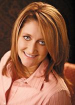
Andrea L. Hegedus owns Great Lakes Smile Design Studio. She can be reached at andrea@glsmiledesign.com.
3D Systems Garners FDA Clearance for Multi-material, Monolithic Jetted Denture Solution
September 17th 2024The company’s unique multi-material, single-piece dentures are designed to offer a combination of distinctive break resistance and outstanding esthetics for enhanced patient experience, and Glidewell labs are currently implementing the solution.