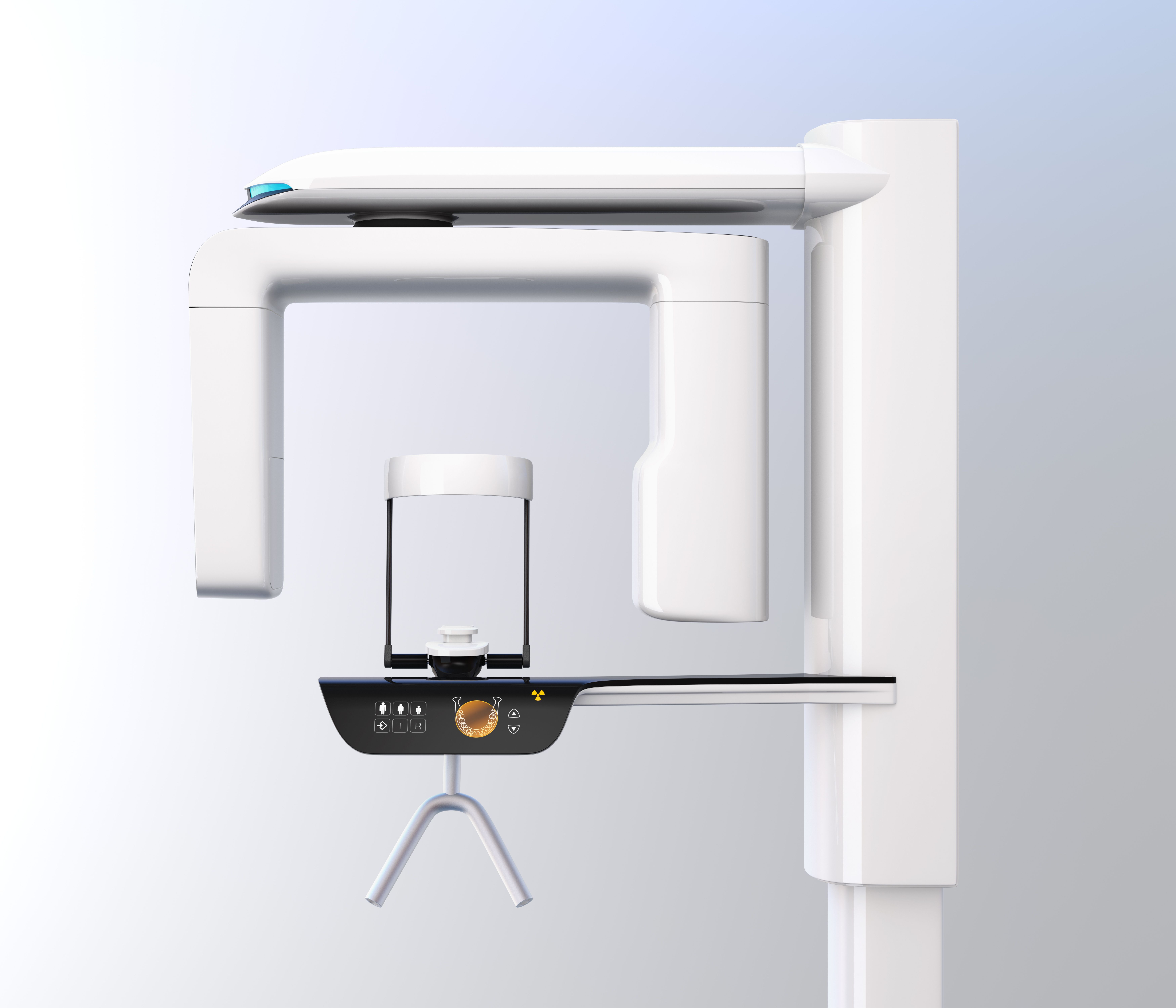
Using Cone Beam Computed Tomography in Endo Cases
3D Imaging and Endodontics – How, when, and why to use cone beam computed tomography in endo cases.
By chesky / stock.adobe.com

Although a map proves useful in getting you where you want to go, it pales in comparison to a globe for determining the relative location of different places or destinations. A map may be better than nothing, but when it comes down to it, a 2D representation of a 3D structure cannot provide an accurate depiction.
As well as applying to our mental image of latitude and longitude, this geometric configuration can skew our interpretation of images of complex anatomy. For a long time, endodontic radiographic assessments were limited to panoramic or intraoral x-rays, which, due to a lack of 3D precision, often make it hard to accurately visualize dentition. For example, a periapical radiograph can be distorted due to superimposed anatomical structures (including the zygomatic arch, maxillary sinus, and roots), making it difficult to get a clear view of root canal anatomy. And, for precision procedures such as the treatment of root canal systems, this lack of clarity can lead to treatment failure. However, newer imaging technology has provided a solution.
According to a joint statement from the American Association of Endodontists (AAE) and the American Academy of Oral and Maxillofacial Radiology (AAOMR) in 2015, cone beam computed tomography (CBCT) has presented a groundbreaking advancement for endodontics. This is due to its ability to visualize the dentition and the relationship of anatomical structures.1
“CBCT has been hugely beneficial to endodontics,” says Dr Richard Mounce, an endodontist practicing in Pacific City, Oregon. “Much of the guesswork about canal location, numbers, curvature, optimal access location, bone levels, pathology, possible nonendodontic lesions, etc, has been eliminated or aided through application of CBCT. In my view, having access to CBCT is the standard of practice, and in certain clinical situations it is the legal standard of care.”
The AAE-AAOMR statement reiterates that small field-of-view (FOV) CBCT images are the image of choice if unusual canal morphology is suspected. These images can be invaluable to diagnosis as they allow clinicians to assess the relationship of teeth to other vital structures, assess traumatic dental injuries such as fracture or resorptive lesions and pathology, and aid in treatment planning for nonsurgical retreatment or surgical endodontics. CBCT is particularly ideal in areas subject to anatomic noise or in cases with symptoms that are difficult to localize, as it can better pinpoint the side of the endodontic issue.
“CBCT imaging has become an essential part of the endodontic world,” says Dr Rebekah Lucier-Pryles, an endodontist in White River Junction, Vermont, and cofounder of Pulp Nonfiction Endodontics. “2D images, like periapical or bitewing radiographs, are limited in their detection ability due to structural overlap (referred to as anatomic noise). 3D imaging removes this overlap and allows clinicians to visualize all areas of the tooth independently.”
These CBCT images are also valuable in retreatment cases, and the AAE and AAOMR believe that previously endodontically treated teeth warrant CBCT scans. According to the organizations, limited FOV CBCT is the imaging modality of choice when examining issues of nonhealing in previous endodontic cases or when assessing endodontic treatment complications, as it can determine what follow-up treatment should occur.1
CBCT has myriad benefits, but like any technology it also has limitations. This makes it critical that clinicians understand when its use is appropriate.
“Opinions differ,” explains Dr Mounce. “Some endodontists take a CBCT on every patient, others when they feel it’s needed for complex anatomy, surgery, retreatment, or resorption cases, etc. I have no hesitations about taking scans, but I personally don’t take a scan on every case. That said, I don’t have any [problems] with clinicians who do.”
The primary concern with CBCT imaging is a possible higher radiation dose for the patient. The AAE and AAOMR recommend that CBCT should not be used routinely for endodontic diagnosis or screening purposes if there are no clinical signs or symptoms that indicate a necessity. Likewise, the organizations state that CBCT imaging should only be used when the imaging needs cannot be met by lower-dose 2D radiography.1
“CBCT should be used only when the patient’s history and a clinical examination demonstrate that the benefits to the patient outweigh the potential risks,” the associations state. “The use of limited FOV CBCT should be considered on a case-by-case basis, with due consideration given to the risks and benefits of exposing the patient to ionizing radiation, the patient’s history, clinical findings, and preexisting radiographs so that superior treatment can be provided to the general public in need of endodontic care.”
In addition to the risk of higher radiation doses, CBCT has other limitations. High levels of noise and scatter and the potential for artifact generation can throw off a clinician who isn’t accustomed to reading CBCT images.2 For clinicians who aren’t comfortable using CBCT, Dr Mounce recommends substituting a surgical microscope. He believes that the lack of both CBCT and a microscope can be of great detriment to the clinician.
“The surgical microscope is a critical piece of equipment and vastly superior to loupes,” he says. “If you don’t have a CBCT and don’t have a microscope, it is exceedingly difficult to envision that a dentist can practice at the same level as a specialist without the microscope. Without a microscope, you simply don’t have visual and tactile control. The double whammy of not having CBCT and/or a microscope is the harbinger of problems.”
Although CBCT presents some challenges that may sway a doctor’s decision to forgo the technology, its increasing availability and expanded applications are making the tool more valuable, despite its current drawbacks. Findings from recent research show that after reviewing CBCT images, many clinicians often change their previously proposed treatment plans because the images so effectively eliminate the concerns over distortion that accompany conventional 2D radiography.3 And, when used as indicated, this 3D imaging can provide clinicians with critical information that allows them to treat cases that may have previously been deemed untreatable.
“CBCT imaging has changed the face of endodontic care, both in its ability to diagnose obscure pathology but also in our ability to treat cases,” Dr Lucier-Pryles says. “Our ability as clinicians to diagnose resorptive dental diseases and to evaluate fracture pathology with CBCT technology has significantly improved, and as a result our treatment strategies are more appropriate.”