Tech Tuesday: Photoacoustic Imaging May Replace the Probe
Technology is quickly transforming dentistry. From digital record-keeping to machines that seem straight out of a SciFi feature, the 21st century has gifted the medical industry some amazing devices. Dentists are especially proactive about adopting new technology in their practices, so DMD is bringing you a weekly spotlight on some of the best products on the market. Continue below to read about this week's focus, photoacoustic imaging.

Using ink from cuddle fish as a contrast agent, researchers were able to highlight inflamed tissue.
Checking patients’ gums for damage can be a traumatic or painful experience for someone who has inflamed or otherwise damaged periodontal tissue. A new study claims it found a way to relieve that pressure by using a new type of ultrasound device.
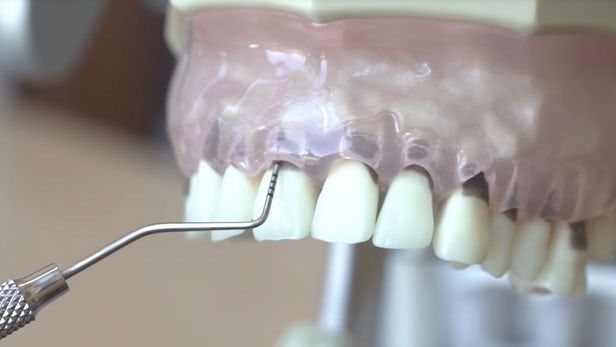
Periodontal probing is the practice of checking on patients’ gums by pressing against the tissue with a small metal rod. Generally, the probe does not damage healthy gums, but if a patient has inflamed tissue, the probe could cause bleeding or actually poke a hole into the gum.
While this method can be painful, it is still the primary way dentists test periodontal tissue for issues. A team of researchers at the University of California-San Diego and UC-Irvine may have found a less damaging method of checking structural integrity and finding issues.
In a study published earlier this month, the team outlines a new method using photoacoustic imaging and the results of their initial testing.
Photoacoustic imaging is the method of using brief laser pulses to light up a tissue sample. The tissues heat up, and are then checked with ultrasonic-frequency sound waves. The result allows the doctor to clearly find damage in tissue structures or compare them to normal samples.
The University of California team tested this method on 39 teeth acquired from pigs, inducing excess damage in 12 of them. Cuttlefish ink was used to highlight pockets and parts of the tissue that were undamaged or inflamed, acting as a contrast agent.
The same teeth were also checked with traditional periodontal probes, according to the study.
The study noted that “there were statistically significant differences between the [two] measurement approaches for” the cheek, tongue, and side of the mouth, but not the middle of the mouth area.
In three out of four areas, the probe and the imaging device found different results from each other.
The imaging technology was better able to look at the tissue than the probe was, covering the entire object being studied. The probe, in contrast, can only be used on certain parts of the area.
The nature of photoacoustic imaging also means it was more precise than the probe could be.
The results indicated that the bias values were less than 25 mm, which indicates a low amount of bias. In other words, the results are expected to be accurate and not just the result of hopeful thinking on the part of the study’s authors.
There was also a low amount of variation between five replicated tests.
In essence, photoacoustic imaging is a potentially less invasive, more accurate method of checking gum tissue for damage or disease. While more work must be done to confirm these results, including testing in humans, the initial results are promising.
“This report is the first to use photoacoustic imaging for probing depth measurements with potential implications to the dental field, including tools for automated dental examinations or noninvasive examinations,” the authors of the study said.
Discover more Dentist’s Money Digest® news here.
RELATED: More Coverage on
- Recognizing Body Dysmorphic Disorder in Cosmetic Dentistry
- Balancing Investments and Student Loans After Dental School
- The Dream of Independent Dentistry is Alive and Well
ACTIVA BioACTIVE Bulk Flow Marks Pulpdent’s First Major Product Release in 4 Years
December 12th 2024Next-generation bulk-fill dental restorative raises the standard of care for bulk-fill procedures by providing natural remineralization support, while also overcoming current bulk-fill limitations.