Enhancing patient communication with DEXIS CariVu
One dentist’s take on how CariVu helps patients better understand their treatment.

They squint. They nod. They say all the right things. But as their dentist, you know your patients do not always understand what you are showing them on the X-ray, and sometimes not even what you are showing them on the intraoral scan. Worst of all, you know patients don’t know why the treatment you are recommending for them is the best course of action for their dental health.
Patient acceptance of treatment plans depends on many factors, and you cannot control all of them. However, you can control what your patients understand about the current state of their dental health. It doesn’t require a dental degree to understand it either. As many a clinician who has invested in technology knows, seeing is believing. When you can show patients pictures of their teeth and blow it up on a screen for them to see magnified, patients will have the information they need to make a sound decision about their dental care.
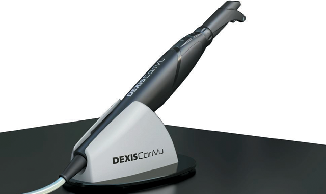
Many technologies help communicate this information in new ways-digital radiographs, intraoral cameras and even laser diodes that emit light to give a digital readout that denotes decay. However, in 2014, DEXIS introduced CariVuTM, a caries detection system that specializes in helping dental professionals show patients what they need to know.
Related reading: Using DEXIS Platinum digital sensors and DEXIS CariVu
DEXIS describes CariVu as a “compact, portable caries detection device that uses patented transillumination technology to support the identification of occlusal, interproximal and recurrent carious lesions and cracks.” But for Dr. Angela Boehler-Homoky, who has been practicing for 23 years and is based in Port Charlotte, Florida, CariVu is a new way for her to communicate what she wants her patients to know about the state of their teeth in a way that they can see for themselves.
We spoke to Dr. Boehler-Homoky about her experience with CariVu. Here’s what she had to say.
What were you using before you got CariVu, and why did you start using it?
I was first using film radiography and then digital radiography. Then I started using intraoral cameras with that. I was looking to purchase a new intraoral camera and learned there was something new also from DEXIS, the CariVu.
What were some of the things you liked about CariVu that were different from what you were using?
I like anything I can show on the screen to my patients so they can see and understand. With some radiographs, I felt my patients were taking my word for it. I’d say, ‘See that spot right there?’ and they’d go, ‘Yeah…I guess.’ I always tell people, ‘This is your body and your health. I want you to understand why we’re doing things and to be included in the decision-making process.’
With digital radiography, you can blow it up and change contrast and try to show them those carious lesions. But many times, they’ll point to a filling. So, I still never felt like they really knew. With intraoral cameras, I thought, ‘Ok great, this is really going to show the patients.’ But I’m finding that with intraoral cameras, when patients see stain, they point to a tooth and say, ‘Oh my gosh, that tooth is horrible!’ And I say, ‘No, that’s an old filling, but it’s doing ok. I’m looking at this.’ And I am pointing to something else, and it’s not the thing that they notice.
With CariVu, they seem to see it, and it differentiates between the normal and not normal teeth. Patients can see why the tooth needs a crown or why it needs a filling.
Trending article: Nominations are open for the 2017 Top 25 Women in Dentistry
So does that increase patient acceptance rates?
Absolutely.
Continue to page two to read more...
What do you like best about using CariVu?
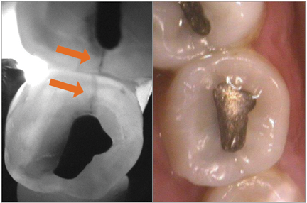
By using CariVuTM, cracks are easily visible to both clinicians and patients.

There are a lot of different things that it does that are unique and useful, but the No. 1 thing I like it for is fractured teeth. I used to call a fractured tooth the mystery tooth. I would say, ‘If a tooth is acting funny in a bunch of different ways, that leads to me to think fracture.’ But I felt like it wasn’t certain and I don’t like that. I like to show a person the crack. Unless it’s an incredibly large fracture, you can’t see fractures on an X-ray at least 99.9 percent of the time. I wear loupes, too, so sometimes I can see them. An intraoral camera sometimes can pick them up. Sometimes there are superficial cracks everywhere. With CariVu, when you suspect the tooth has fractures, you put it on and take an image. I can put that image up on the screen, and the patient can see it just as easily as I can. And the insurance companies can see those cracks as well. I love CariVu for that.
I would think the patient would love that the insurance company can see it.
That’s true. Insurance gets kicked back with, ‘Why are you putting a crown with an occlusal amalgam?’ I send them the CariVu image and say, ‘Because there’s a huge fracture going through it, and the patient is going to break this tooth.’ The insurance company can see it, and everyone agrees. You can’t argue with facts.
Trending article: DEXIS introduces the new DEXcamTM 4 HD intraoral camera
What advice would you have for a clinician struggling with patient acceptance rates or some of the problems you were having?
With intraoral cameras and CariVu, patients get it. I want my patients to trust me, but I also want them to see what I see without having to have a dental degree. And this is the easiest way.
From the patients’ standpoint, they don’t get that much out of the X-ray. They get some information out of the intraoral camera, but then they really see with the CariVu. From the doctor’s side of things, if there was overlap on the bitewing, which happens all the time with crowded teeth, you can miss the decay. But it shows up on CariVu images even more than on an X-ray. There’s no guesswork with the CariVu looking for interproximal decay. It’s dark; it shows up. For a dentist’s eye, there’s no question now.
Is there anything else you want to mention about the system or the integration?
Occlusal decay. Not a lot of devices show occlusal decay well. I have another product I use to check for fissures and things, but I don’t think it does well around existing restorations and sealants, I don’t feel. Sometimes I’ll get a false or high reading around some sealants and some restorations. Or if there’s plaque or calculus, it will give a false reading because that’s not very dense.
On occlusal decay, I don’t like devices that give false readings because now you’ve got information that you can’t utilize. It’s something thrown into the mix to confuse you. With CariVu, there’s no false reading. It can look through composites and sealants and calculus or plaque. It’s just showing the decay that’s there or the restoration of the tooth.
And X-rays don’t really show occlusal decay very well either unless it’s big enough. Most of the time when the occlusal is big enough to see on a radiograph, you can see it visually. So CariVu is good for those smaller ones.
Do you have any tricks from using CariVu that you would share with other clinicians?
A newbie mistake that I see is not drying the teeth out first. If one of my assistants is trying to use it and takes a picture when the teeth are not dried out, we get a nice picture of bubbles. A quick blast of air over those occlusions usually gives you a much easier to read picture for sure.
Read more: DEXIS delivers enhanced CariVu software
Are there any other tips you have picked up along the way?
There is a small learning curve because it’s a different instrument than probably anybody has, as far as putting it on the tooth. But it’s a quick learning curve that only takes one or two tries of practicing on each other. It’s easy to use.
It works alongside DENTRIX easily. When I’m going to use CariVu, if I already have an X-ray or a picture, and most of the time you do, as soon as you put the CariVu in and select what tooth you’re taking a picture of, it pulls up the X-ray in a side-by-side screen. Then, it pulls everything that you’re going to present to the patient or look at for comparison automatically. I know I sound like a commercial, but I’m not trying to. I’m just saying what it is. It makes it easy. It not only integrates, but it also has bells and whistles that make it even better.
How Dentists Can Help Patients Navigate Unforeseen Dental Care
December 12th 2024Practices must equip patients with treatment information and discuss potential financing options before unexpected dental treatments become too big of an obstacle and to help them avoid the risk of more costly and invasive procedures in the future.
Product Bites – January 12, 2024
January 12th 2024The weekly new products podcast from Dental Products Report is back. With a quick look at all of the newest dental product launches, Product Bites makes sure you don't miss the next innovation for your practice. This week's Product Bites podcast features new launches from Videa Health and DentalXChange.