Dental Innovator: How DEXIS helps the dental industry keep its cool
We talk with Carsten Franke and Boaz Munnerlyn of DEXIS about the company's path to success and how it's helping dentistry become "cool." Q: I feel like this Q&A should start with a “Thank you” - DEXIS has provided several outstanding product launches to feature in DPR this year!
We talk with Carsten Franke and Boaz Munnerlyn of DEXIS about the company's path to success and how it's helping dentistry become "cool."
Q: I feel like this Q&A should start with a “Thank you” - DEXIS has provided several outstanding product launches to feature in DPR this year!
A: Thank you very much! You’re right, the launch of DEXIS® Imaging Suite and its companion app for the iPad, DEXIS go®, have been two major product launches in a short period of time. Both are very innovative software solutions and completely indicative of the long line of DEXIS firsts that started when we introduced DEXIS in the US in 1997 - which, coincidentally, was on the cover of DPR...
Q: Tell me more about this history, and what sets DEXIS apart.
A: Our products are clinically relevant, ergonomically designed, simple to learn and easy to use. It all started with the DEXIS Classic sensor in the late ‘90s which made digital X-ray imaging portable and affordable for the first time. Remember, back then we truly revolutionized digital radiography and became the catalyst that drove the adoption rate of this technology in North America! We invented image enhancement tools like ClearVu™ - a remarkable step forward for clinical diagnosis.
In 2000, we were already in conformance with the DICOM Standard (Digital Imaging and Communications in Medicine); years before others even thought about it.
In 2001, we were the first, and for many years the only, digital X-ray system to be accepted into the prestigious ADA Seal Program following an extensive product evaluation.
In 2009, we took the gold standard in digital radiography to a platinum level by refining the design of our PerfectSize™ sensor and creating the DEXIS Platinum® direct-USB sensor, and, thus, eliminating the need for multiple-size sensors and adaptor boxes. And of course, this brings us back to DEXIS Imaging Suite and DEXIS go.
Q: How does the evolution of the DEXIS imaging software and its ability to work with DEXIS go on the iPad fit into the larger pattern of innovation the industry has come to expect from DEXIS?
A: DEXIS Imaging Suite and DEXIS go show our core philosophy of simplicity while incorporating new features that are becoming relevant now. Both programs offer feature-rich yet easy-to-use functionality, a combination that requires much thought and an understanding of what dentists need - and these products are building the platform for exciting future applications.
DEXIS is the leading imaging solution serving general dentists, specialists and the forensic community. We continue to refine our product offerings, and due to these efforts, we have been highly awarded by researchers, well-respected dental publications and the dental community.
We’ve set the bar pretty high for ourselves. The dental community expects us to continue to be a leader - and DEXIS is committed to meet their expectations. We are humbled to receive the 2013 Best Technology Award from Pride Institute for DEXIS Imaging Suite and DEXIS go; it’s an honor that the Institute values the innovation and ingenuity in these new products that can help clinicians on a daily basis.
Q: With two huge developments rolled out in a relatively short period of time, what can we expect next from the R&D team at DEXIS?
A: We created some exceptionally innovative features and tools with our last two products, especially around treatment planning and patient presentation. I cannot comment on unreleased products, but I can tell you that our R&D and Engineering teams are hard at work. And looking at the pipeline in our labs, I’m thrilled about what’s coming.
Q: Our internal survey data indicates that 75% of dentists are using some form of digital imaging in their practice. If you had to make your case to those who are still on the fence about digital radiography, how would you summarize the benefits?
A: Our research shows very similar results. It is great to see that digital radiography has been so widely adopted by now and clinicians and patients alike are enjoying the great benefits of this technology. Our data also indicates that the majority of practices still using film today are actively looking into digital imaging solutions.
And if you really think about it… switching from film to direct digital is a no-brainer. No darkroom, no chemicals to deal with, no ongoing expenses for film as long as you practice, instant images, shorter appointments, more chair time, more patients you can see, large X-ray images you can display on a monitor, TV or iPad, better patient communication and increased treatment plan acceptance. And not to forget… reduced exposure to radiation. Again, it’s a no-brainer.
Q: Some companies have tried to use the iPad platform to be trendy. DEXIS go® seems to be about more than that. What sets it apart from other apps?
A: DEXIS go is not a “trendy” fad. It brings relevance to the patient experience. With all imaging information at their fingertips, the dental office team can now more easily communicate to patients in a compelling way that makes patients feel comfortable and included in the valuable discussion about their oral health care. There’s also the WOW-factor: one of the coolest features in DEXIS go is our Lightbox mode, an homage to dentistry’s past in a very contemporary product.
Q: How does DEXIS go improve the overall workflow for the dentist?
A: With the DEXIS go app, all of the patient’s intraoral and extraoral radiographic and photographic images within DEXIS Imaging Suite can be viewed wirelessly on an iPad. The app fully supports the device’s touch-operation including swiping and pinch-to-zoom. Even ClearVu can be applied. Discussing the treatment plan with the patient can occur anywhere-in operatory, consult room, front office - on an iPad, a communication device that so many people these days are comfortable with.
Q: How does the iPad experience change a patient’s view of treatment planning and his or her oral health?
A: Because of products like DEXIS go, we may be seeing the end of the era of negative attitudes toward the dental office experience. Patients can be included in the discussion of their health care like never before. Dr. Chris Anderson, a proponent of using mainstream technology to further the dentist-patient connection, puts it this way: “The images simply come alive in their hands.” It’s a wonderful way for them to learn. Dentistry has never been “cooler” than it is right now.
The 5-Minute FMX with DEXIS Digital X-ray
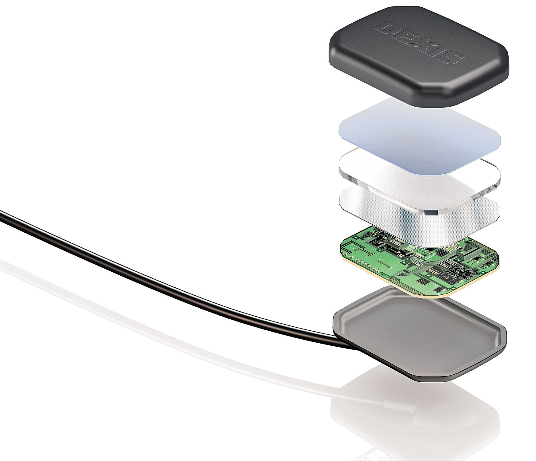
While every digital X-ray system saves time over traditional film, you really need a single-sensor solution to maximize time savings. DEXIS offers the PerfectSize™ Platinum sensor-an ergonomically designed universal sensor that can help keep you moving quickly.
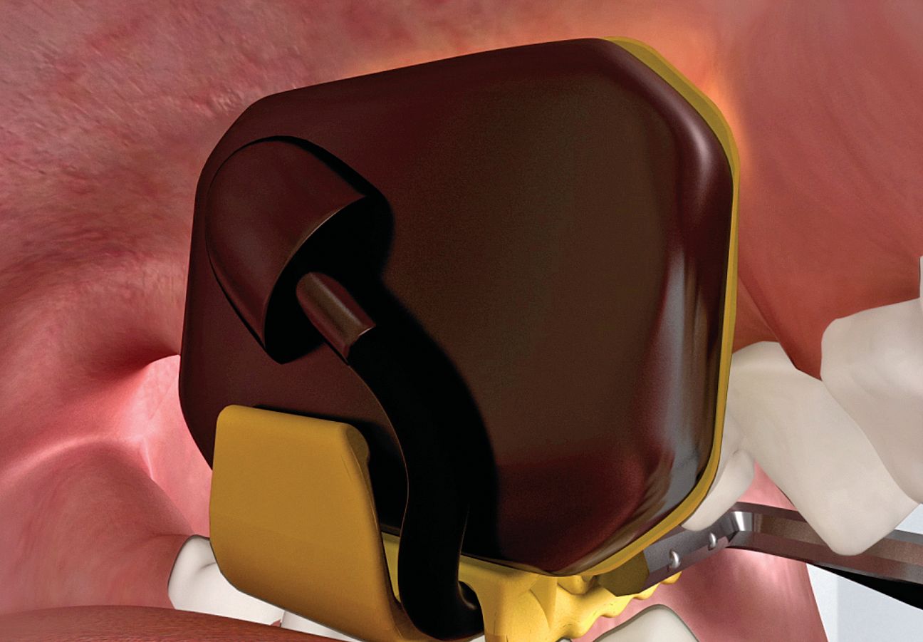
Good design is key: Beveled corners and a smoothly rounded casing allows the clinician to more efficiently move about the mouth with the sensor, even in tough areas such as upper molars and lower premolars, partly because it is more comfortable than square-cornered sensors. Also, more comfort comes from the sensor holder design. Biteblocks seamlessly hug the sensor so as not to produce sharp edges.
Working in conjunction with the sensor and holders, the software does its job instantly. With each capture, the image is automatically saved, dated, tooth numbered, correctly oriented and mounted, and you also gain a preview. You also can set up your own sequence-choose how you want to take X-rays in an order you find most efficient.
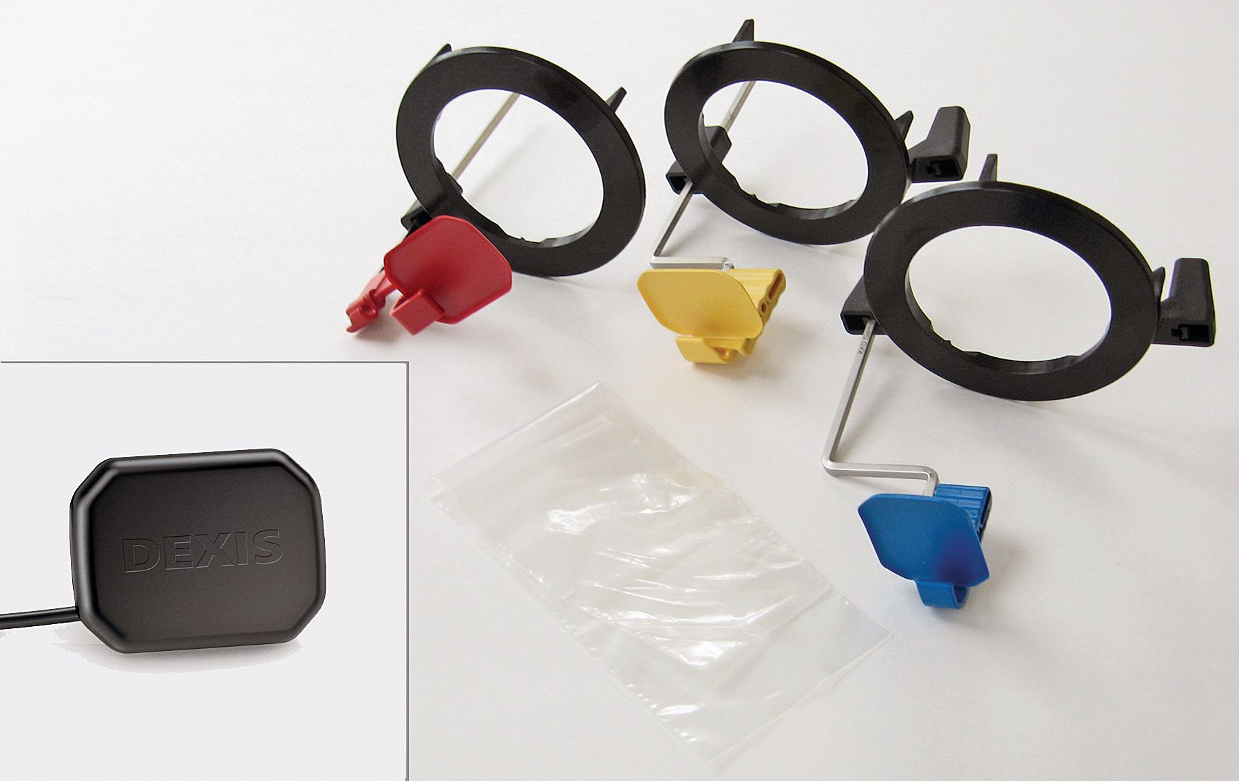
Physical Set-up: Barrier the sensor (not multiple sensors) and assemble the rings, bars, and biteblocks that have been autoclaved per CDC guidelines. Note: DEXIS uses RINN-style holders that are familiar to most dental professionals.
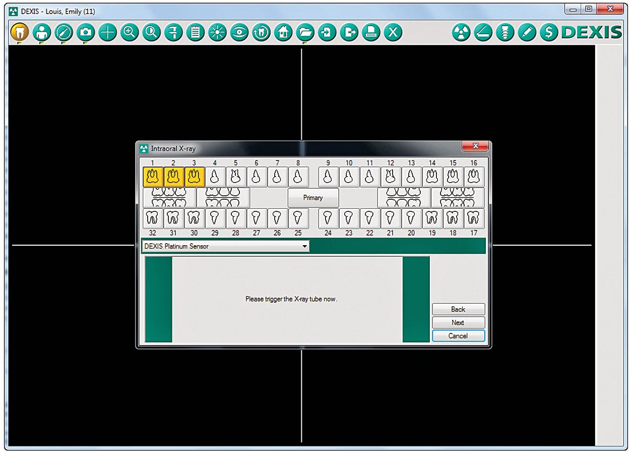
Software Set-up: With the patient intraoral X-ray screen open, click on the X-ray Acquisition screen and then click on Full Mouth. The first area to be captured is highlighted in gold. Here, the default sequence is shown.
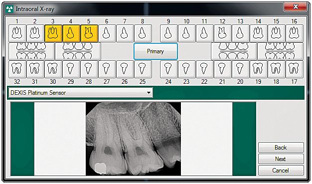
Capturing Images: Position the sensor in the first posterior area and trigger the X-ray tube. You will gain a preview of this image and the software will advance to the next position. Capture the rest of the posterior images using the same biteblock, ring and bar. For adjacent areas (i.e.: UL molar, UL premolar), there is no need to remove the sensor, simply readjust its position. Next, change to the anterior biteblock and capture anterior images.
Note: You have just saved time because you do not need to switch to another sensor and additional biteblocks! Again, for each adjacent area, there is no need to remove the sensor.
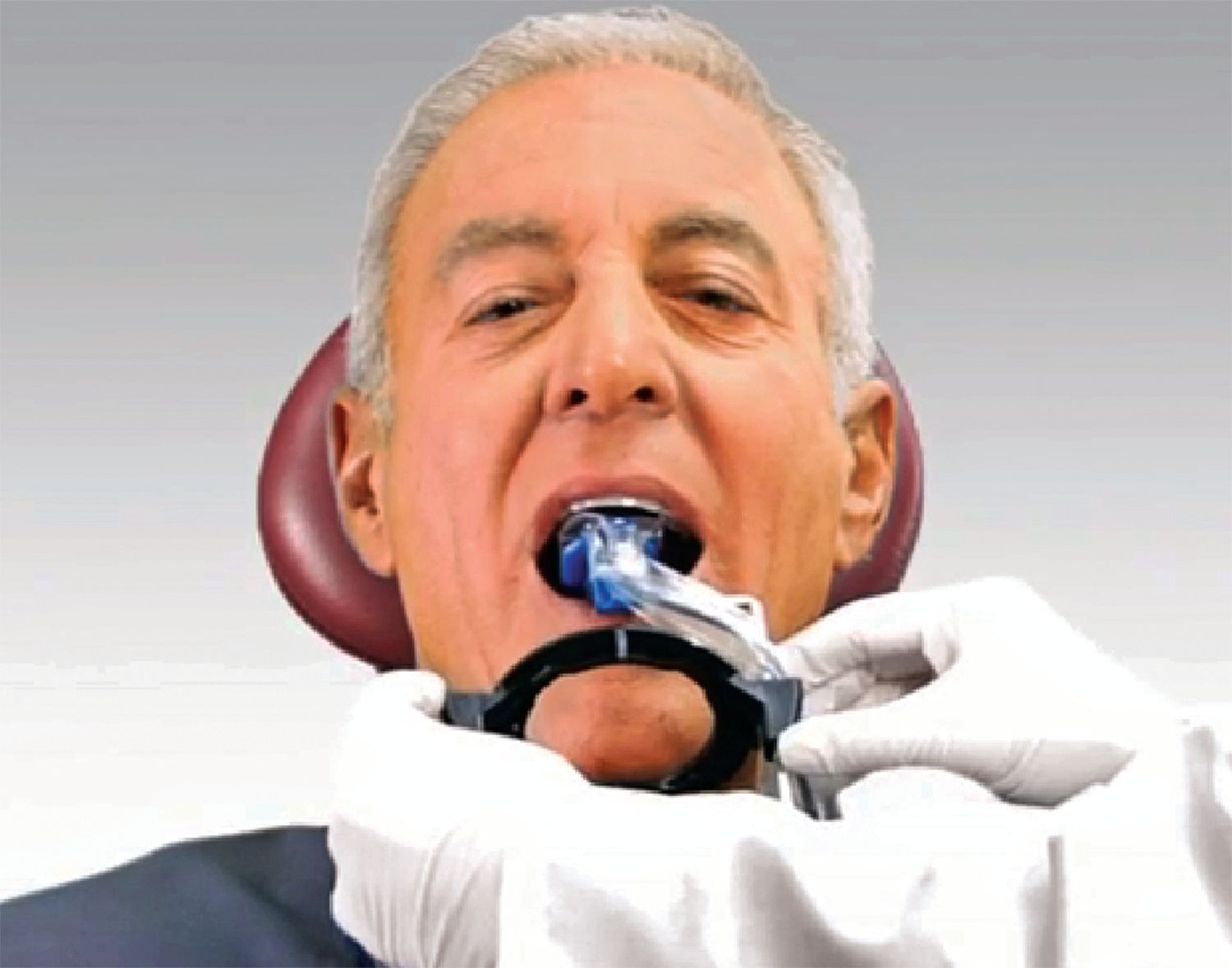
Change to the bitewing holder assembly and capture either horizontal or vertical bitewing X-rays.
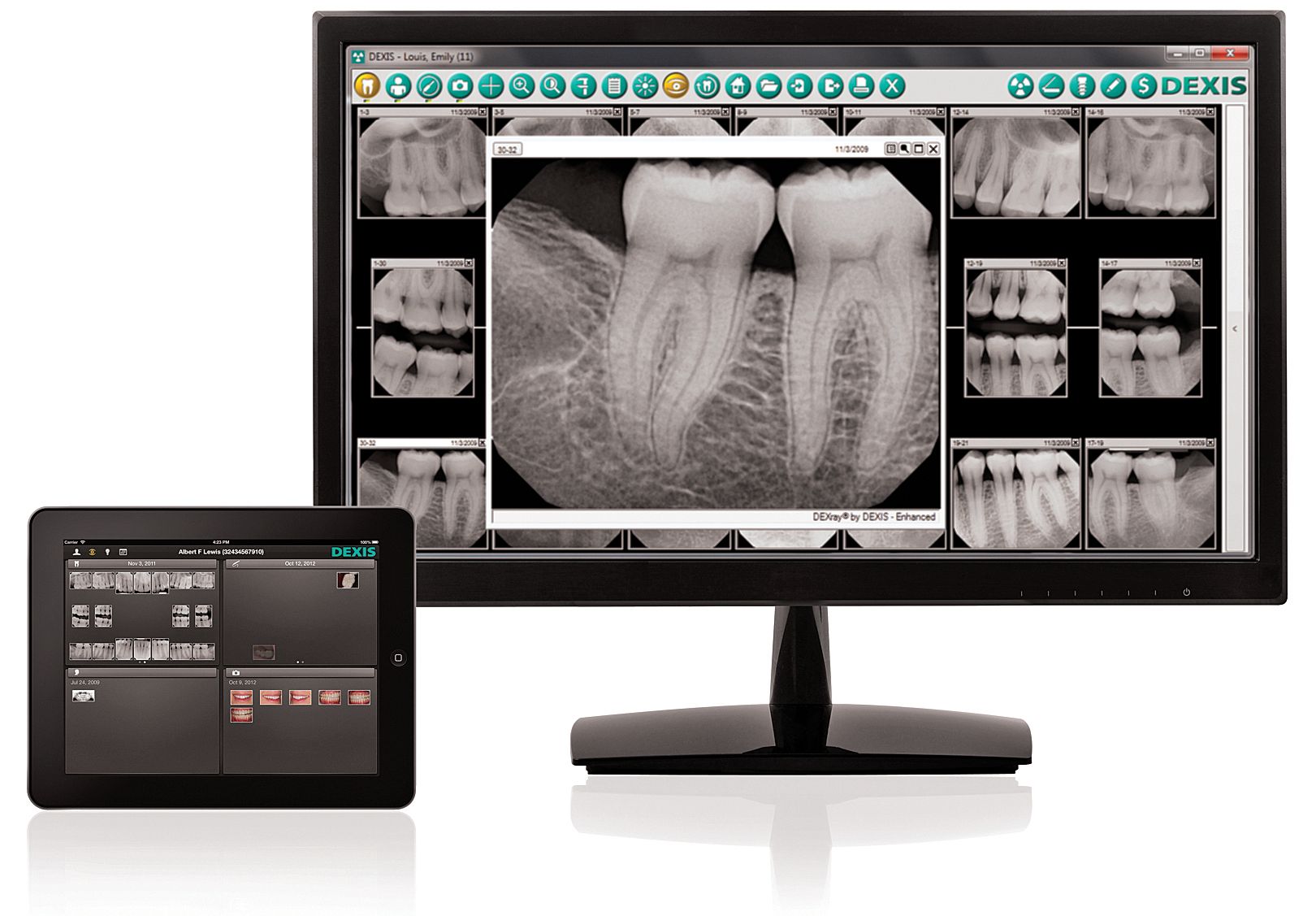
When done, all your images will appear on the screen, already numbered and mounted, immediately ready for diagnosis - in less than 5 minutes! And they are available on your iPad device using the new, free and innovative DEXIS go® app - for DEXIS® Imaging Suite 10.0.5 and higher.
Physical Clean-up: Remove and discard the barrier and use the appropriate cleaner on the sensor. Rinse, bag and autoclave one set of holders (not two sets).
Recap:
• The sensor and biteblock design allow you to move efficiently, quickly and comfortably.
• There’s no switching to different size sensors and their holders.
• You choose your own sequence, the one you are comfortable with.
• There’s less set-up before and clean-up after the procedure.
• The software does the processing and mounting for you.
FEATURED PRODUCTS

This direct-USB digital X-ray solution features the PerfectSize™ Sensor, allowing clinicians to take vertical and horizontal bitewings as well as all periapicals with one sensor; a gold-plated USB connector; the proprietary WiseAngle™ Cable Exit that provides the cable flexibility to reduce stress and increase reliability; and TrueComfort™ Design that offers a slim profile, four beveled corners and rounded casing for patient comfort and precise placement. PureImage™ technology offers optimal image quality.
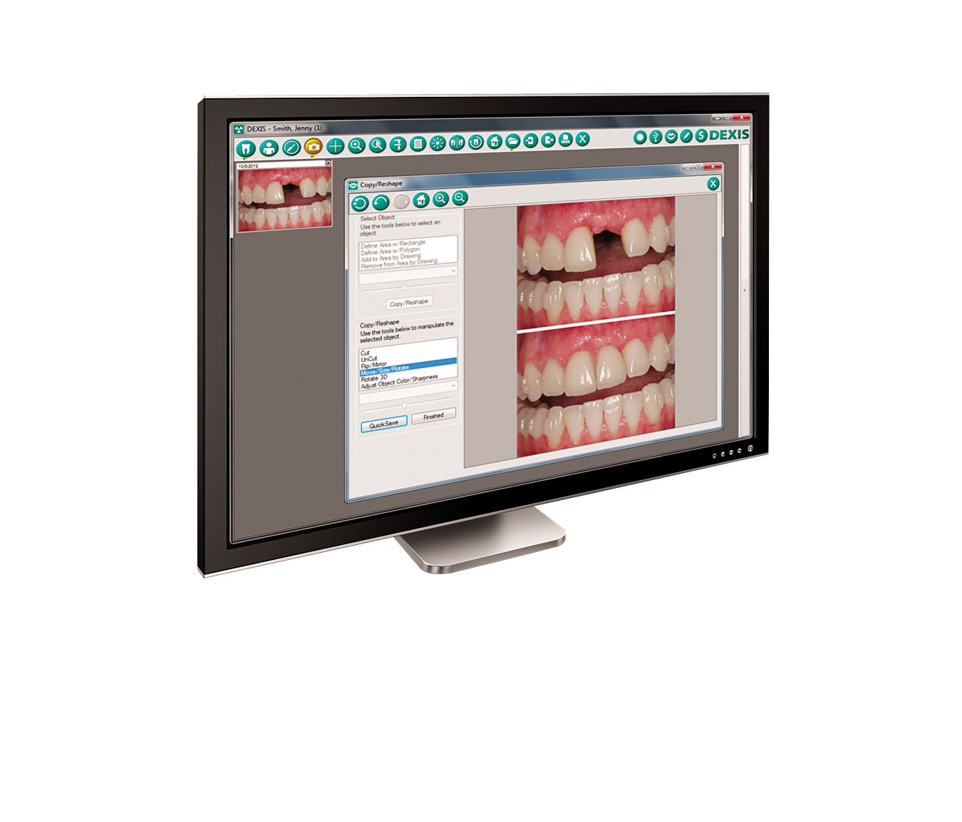
Providing progressive software architecture and adding new features, DEXIS Imaging Suite 10.0.5 includes a cosmetic imaging module, expanded video capabilities, an enhanced implant planning module and integration with select 3D products. The cosmetic module permits clinicians to plan, simulate and present full cosmetic procedures and tooth whitening treatments in just minutes. The software integrates 3D scanners from i-CAT®, Gendex®, Instrumentarium®, and Soredex® - allowing users to manage patient data and 3D images directly from the DEXIS application.
ACTIVA BioACTIVE Bulk Flow Marks Pulpdent’s First Major Product Release in 4 Years
December 12th 2024Next-generation bulk-fill dental restorative raises the standard of care for bulk-fill procedures by providing natural remineralization support, while also overcoming current bulk-fill limitations.