How to use transitional bonding for interdisciplinary care
The growth of multiple options to serve patients’ esthetic and functional needs and desires requires mastery of disciplines that usually necessitate a coordinated and collaborative team approach with multiple specialties.
The growth of multiple options to serve patients’ esthetic and functional needs and desires requires mastery of disciplines that usually necessitate a coordinated and collaborative team approach with multiple specialties.
Often, the information is in the form of the traditional database of radiographs, photos, models and diagnostic wax ups, as well as new 3D digital technologies such as cone beam (DICOM files) and impressions (STL files). More importantly, creating a shared vision of the final esthetic communicates infinite details of the patient’s hopes and dreams and presents higher accountabilities between the restorative, orthodontic and surgical partners (aka the three Rs: relocate, replace and restore).
Fortunately, with this high level of expectations, there are innovative, conservative and dynamic methods that keep everyone on the same page and satisfy a patient’s emotional appearance needs during interdisciplinary care that may take several months or years.
This article will demonstrate how the general dentist can “quarterback” and design success as an architect and an artist.
Related reading: GC America unveils NEW GC FUJIROCK line in bulk packaging
Case study
A 35-year-old female patient with a history of unsuccessful surgical exposure/orthodontics eruption and subsequent extraction and bridge reported to our office with a misplaced implant at tooth position No. 11. Because of the compromised bone volume caused by the rotated first bicuspid, the surgeon in South America was unable to access a proper location for the implant three dimensionally, which compromised the trajectory toward the labial, as well as the attached gingiva. With the situation creating an unacceptable esthetic, biologic and functional risk, the patient accepted a long-range treatment plan of orthodontic and surgical site development prior to placing an implant. This would be followed by restorative care with an implant-supported crown using custom zirconia abutment for No. 11 and porcelain restoration on adjacent teeth to create an ideal smile enhancement.
Check out the following video to view the step-by-step case:
Related reading: In memory: Richard Atanay of GC America
Continue reading about the procedure on Page 2 ...
To be able to communicate our future goals and parameters to the orthodontist and periodontist, records were gathered and shared, including Digital Smile Design to road map the journey. The sequencing of care was “brainstormed” in the following phases:
- Full orthodontic realignment of the teeth, in addition to creating adequate space for increased bone volume and implant placement. The pre-existing transitional bridge was to be split to allow for distal rotation of tooth No. 12 to create more optimal intradental space.
- Esthetic verification of the ideal placement of tooth No. 11 with an updated provisional restoration, as well as CBCT analysis for surgical planning.
- Soft tissue and block osseous grafting prior to a tapered implant placement with a surgical guide.
- Restoration as listed above and occlusal maintenance with a retainer and protective appliance.
Related reading: GC America introduces GC Initial MC Classic line
1. Following two years of orthodontic therapy, a space was created between Nos. 10 and 12, necessitating a more ideal wax up to plan a provisional that diagrammed pink/white balance and position of the missing canine.
2. A lined silicon matrix was created to help duplicate this intraorally (Sil-Tech putty, Ivoclar Vivadent). The patient was given the opportunity to home whiten using her Essix retainers. Also the gingival shades were assessed prior to anesthesia of the tissues, which would affect blood flow and color in the area of treatment.
3. After local anesthesia, the length of tooth No. 10 was enhanced using an all-tissue Erbium laser (BIOLASE) with soft tissue settings of 2 watts/20% water/20% air.
4. The silicon matrix was filled with a Bisacryl Luxatemp Ultra B1 material (DMG America) with an embedded custom cut and resin-coated strip of 3 mm wide Ribbond (Ribbond) that added structural reinforcement to prevent cracks from propagating.
5. After full curing of the restoration, it was shaped and equilibrated. Some excess material was cut back facially to allow for layering of GRADIA® gum shades composite material (GC America).
6. Using GRADIA Pink instructions from the manufacturer,3 we roughened the resin surface by sandblasting with 27 micron aluminum oxide using a PrepStart (Danville Engineering).
7. After full curing of the restoration, it was shaped and equilibrated. Some excess material was cut back facially to allow for layering of GRADIA® gum shades composite material (GC America).
8. A coat of GC COMPOSITE PRIMER was placed on the roughened surface to wet the Bisacryl and promote adhesion and was light cured for one minute. Finally, two colors were layered anatomically and separately onto the surface. The initial application (shade G23) was placed more apically and corresponds to a darker, unattached gingival tissue. The final layer (shade G21) was blended into the G23 and sculpted to match adjacent attached gingiva.
Video: GC America's G-CEM LinkAce tackles the common challenge of indirect restorative treatments
Read the rest of the steps on Page 3 ...
9. Unfilled resin helped the application of this material. This was smoothed and shaped to mirror the corresponding anatomy including the papillae.
10. The tooth portion of the restoration was tinted with a thin layer of ochre shade to blend with enamel on tooth No. 9 using Kolor Plus (Kerr).

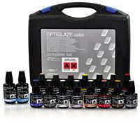
11. The provisional was completely sealed with OPTIGLAZE™ (GC America) to enhance physical and optical durability. Afterward, it was carefully cemented using GC TEMP ADVANTAGE®, which contains fluoride, potassium nitrate and chlorhexidine for pulpal protection.
12. Following this procedure, the incisal edges, which had been worn down from excessive function, were enhanced to promote better esthetics. Given that this was an additive technique, light beveling of the enamel surface was followed by Prepstart microabrasion (rinsed by Ultradent Products’ Consepsis) and 37 percent phosphoric acid (washed thoroughly away by water and lightly dried to allow some moisture with no desiccation of the surface). Placement of a multipurpose resin and curing for a minimum of 30 seconds will help prime the surface for the restorative material.
Video: GC America explains the innovative initiator system of new G-CEM LinkAce
13. Using a compule delivery system, small amounts of shade BW KALORE™ composite (GC America) were applied and sculpted to add a more youthful look to the central incisors and help the incisal plane coincide better with lips and face.
14. After polishing with multifluted carbide finishing burs, as well as abrasive discs and aluminum oxide paste, the patient was very pleased with her rejuvenated appearance.
15. After taking a CBCT of the area, it was easy for the periodontist to visualize the “triangle of bone” and collaborate with us and our patient on the implant position relative to the ideally placed pontic on the provisional bridge and the need for osseous/soft tissue augmentation.4
Phases three and four have yet to be completed. This may take another 12 months due the necessity of optimized healing from grafting and implant surgery before the restorative phase can begin. Fortunately, the patient will be able to feel comfortable with her appearance.
Conclusion
Careful treatment planning of complex cases requires a collaborative approach that allows the interdisciplinary team to share information and create optimized results by not only sharing information to blueprint plans for excellence but also to allow the patient to participate in his or her care. When there are esthetic considerations, having input from specialists, lab technicians and the patient will allow the team to move forward in tandem so everyone is on the same page. Using transitional bonding offers a modality to conservatively communicate esthetic goals, test drive the blueprint and give the patient a chance to enjoy a nicer appearance during multiple phases of care that often take many months to complete. Fortunately, we have a wide palette of colors to customize and balance pink and white considerations.
Review: GC America's Fuji TEMP LT has excellent handling, low film thickness
About the author
Hugh D. Flax received his DDS degree from Emory University and completed his residency at LSU Dental School. He has been a member of the American Academy of Cosmetic Dentistry since 1994 and became accredited in 1997. He was president of the Atlanta Chapter of the AACD (1996-1998) and founded the Georgia Academy of Cosmetic Dentistry in 2007. Dr. Flax served for two years as cochair of the Conference Advisory Committee for 2003 AACD Scientific Session in Orlando. He has been the chairman of the AACD Private Education Advisory Council, the 2008 AACD Meeting and the 2010 AACD European Meeting. He was a member of the AACD Board of Directors for more than eight years. Outside of the AACD, Dr. Flax is a member of the ADA, AGD, ALD, AAID, ICOI, Catapult Elite and the L.D. Pankey Alumni Association. He is also a certified fellow with World Clinical Laser Institute and a fellow of both the International Academy of Dentofacial Therapeutics and ICOI. Dr. Flax practices full time in Atlanta, focusing on functional appearance-related conditions and advanced laser dentistry, as well as writing and lecturing on helping others do the same for their own practices. In 2003, he was WSB-TV.com’s expert on cosmetic dentistry and also appeared on FOX News. He has been the expert dentist on the “Meet the Products” television show and H2O Magazine. Lastly, he and his practice have been featured in many women’s magazines.