Bench mastery: Complete implant maxillary and mandibular reconstruction
Luke S. Kahng, CDT, goes through a step by step technique on how to complete a implant maxillary and mandibular reconstruction.

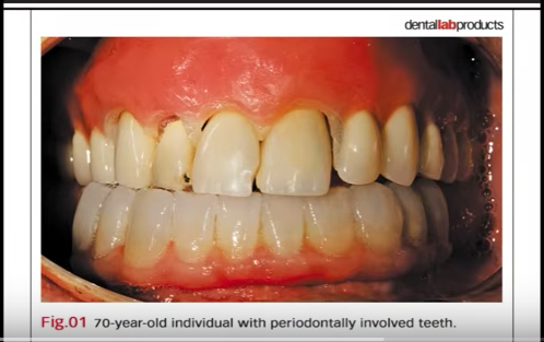
In this case study, a 70-year-old individual with advanced periodontally involved teeth having class three mobility presented for comprehensive implant restorations.
He had few remaining teeth, necessitating extractions. His desire was for fixed restorations, not removable prosthetics. The patient was in good health with no medical contra-indications for the surgical/prosthetic endeavors he was about to undergo.
Case study
1. After a necessary prosthetic workup, which consisted of diagnostic models and records, according to standard prosthetic protocol, treatment dentures were constructed. The maxillary arch had sufficient bone volume and adequate width and vertical height for implant protocol without grafting. A cone beam CT was taken with a treatment denture with radio-opaque markers for designing a stereolithographical stent for a mucosal-born surgical guide for the placement of seven maxillary implants. Our oral surgeon placed the implants and, after sufficient healing time, the impression of the maxillary implants using open tray technique.
Related reading: Bench mastery: How to foresee the final outcome of a dental restoration case
2. The healing was uneventful. The patient followed recommended care for plaque removal around the healing abutment and cleaning of the denture. His hygiene was excellent. Four months later, after ascertaining integration, the technician proceeded with the sequence needed for completion of the maxillary arch.
3. In order to establish a stable centric relation record, it is necessary to utilize a treatment denture setup that is stabilized on two implants. This denture is screwed into the two implants for stabilizing of the maxillary rim to establish centric relation record. After the multiple sequence consisting of framework and bisque bake try-in using the lab protocol, the maxillary prosthesis was completed. It was decided to divide the maxillary arch into two units.
Check out the following video to view the step-by-step case:
4. Four implants with the final right segment were completed.
Bench mastery: Custom shade matching with an anterior implant case
5. This article would not be complete without describing the protocol and procedures in the reconstruction of the mandibular arch. There are several steps required for understanding of aberrant bone level. This image shows few remaining mandibular teeth that required extraction. Notice the bone level in the mandibular anteriors was not in harmony with the mandibular bilateral posterior ridges. The remaining bone was thin from a labial lingual view. Bone supported steriolithographical surgical guides Fig. 10 were fabricated for bone reduction and implant placement. It was necessary to use the bone reduction guide seen here to perform a horizontal osteotomy to level the bone in the mandibular anterior to create a harmonious arch. After completion, note the surgical guide was used after the anterior reduction was accomplished, during which four implants of wide diameter and 15 mm in length were surgically placed. It was determined that stability using wide body implants and 15 mm length with 3 mm of circumferential bone around each implant, accomplished with a horizontal osteomtomy, was adequate to accommodate a fixed restoration.
6. The complete review of all the necessary surgical stents required to achieve results discussed above.
Video: Bench mastery: Treating the dental-phobic patient
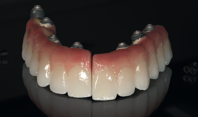
7. The restorations with pink gingival color porcelain support were completed, maxillary and mandibular arch, as illustrated. The design of the prosthesis was in accordance with the patient’s ability to maintain oral health. All openings for floss were created from the lingual for oral hygiene maintenance by the patient.
8. The patient was pleased with the fixed implant prosthesis.
